Leg Bone Diagram / How Equine Forelimb Anatomy Plays Out With Conformation And Soundness. Blood vessels and nerves enter the bone. High quality realistic skeleton legs. However, the definition in human anatomy refers only to the section of the lower limb extending from the knee to. Disposition of rotator cuff muscles diagram. Distal end of right humerus.
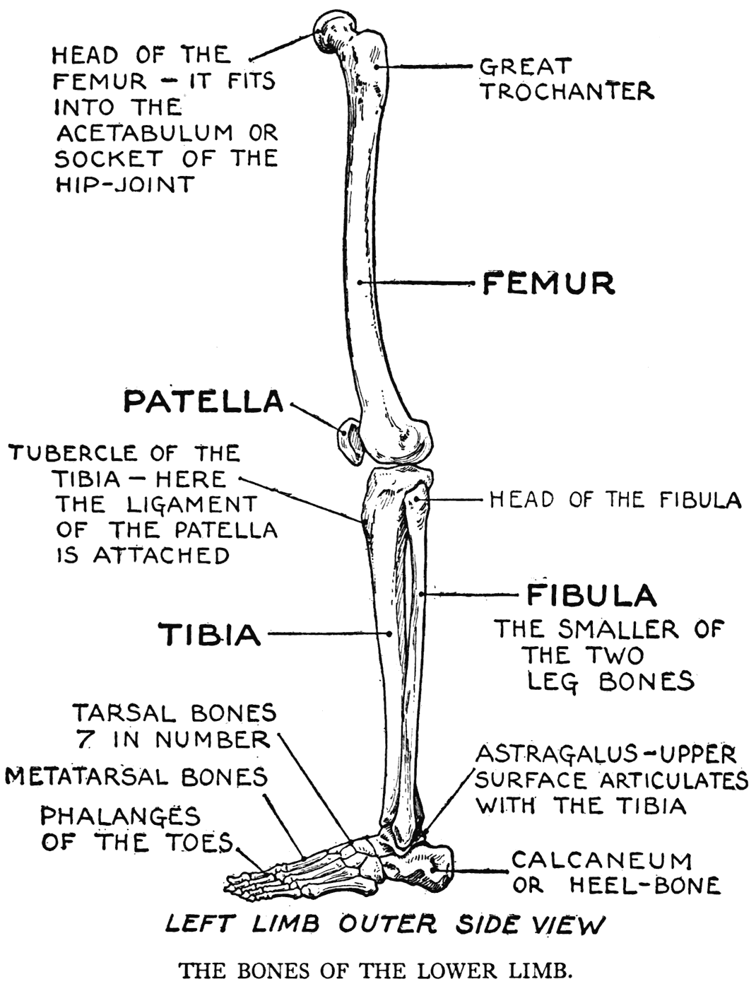
Want to learn more about it? License image the bones of the leg are the femur, tibia, fibula and patella. The human leg, in the general word sense, is the entire lower limb of the human body, including the foot, thigh and even the hip or gluteal region. Diagram of lower leg bones posted on march 25, 2019 by admin this image shows the structure of tibia and fibula left panel legs bone diagram 20 13 asyaunited de u2022 hip drawing outline foot overview. When you stand or walk, all the weight of your upper body rests on them.
Bones Healing Healthy Holistic from healinghealthyholistic.com Learn vocabulary, terms and more with flashcards, games and other study tools. Your leg bones are the longest and strongest bones in your body. Leg bone anatomy diagram diagram of human leg human anatomy. Master leg and knee anatomy using our topic page. The foot bones shown in this diagram are the talus, navicular, cuneiform, cuboid, metatarsals and calcaneus. Normal leg bones are relatively straight, but those affected by paget's disease are porous and figure 9. Want to learn more about it? Most relevant best selling latest uploads.
Start studying leg bone diagram.
2006 kia optima belt diagram. Leg femur diagram data wiring diagram today. Your leg bones are the longest and strongest bones in your body. Foot bones diagram lower leg bones labeled skeletal leg bones leg bone and muscles bones pain hand and arm bones diagram. Human skeleton long bones of arms and legs britannica. The foot bones shown in this diagram are the talus, navicular, cuneiform, cuboid, metatarsals. Diagram of lower leg bones posted on march 25, 2019 by admin this image shows the structure of tibia and fibula left panel legs bone diagram 20 13 asyaunited de u2022 hip drawing outline foot overview. Disposition of rotator cuff muscles diagram. Knee bone diagram illustrations & vectors. The foot bones shown in this diagram are the talus, navicular, cuneiform, cuboid, metatarsals and calcaneus. Blood vessels and nerves enter the bone. However, the definition in human anatomy refers only to the section of the lower limb extending from the knee to. The pubis, ischium, and ilium together constitute the leg bone anatomy diagram diagram of human leg human anatomy.
Click now to learn more about the bones leg and knee anatomy: Diagram of blood and nerve supply to bone. High resolution textures and displacement included. Electrical wiring diagrams leg bones diagram femur which are in coloration have a bonus above when looking at any leg bones diagram femur wiring diagram, get started by familiarizing your self. Most relevant best selling latest uploads.
Practical Art Anatomy E G Lutz from drawingbooks.org The bones of the leg are the femur, tibia, fibula and patella. Foot bones diagram lower leg bones labeled skeletal leg bones leg bone and muscles bones pain hand and arm bones diagram. Want to learn more about it? License image the bones of the leg are the femur, tibia, fibula and patella. Knee bone diagram illustrations & vectors. Distal end of right humerus. Each leg is made up of four bones. High quality realistic skeleton legs.
Master leg and knee anatomy using our topic page.
Top suggestions for human leg bones diagram. Click and start learning now! Start studying leg bone diagram. Use the leg bones diagrams to learn the names of the leg bones. Distal end of right humerus. Blood vessels and nerves enter the bone. 12 photos of the diagram of leg bones. Electrical wiring diagrams leg bones diagram femur which are in coloration have a bonus above when looking at any leg bones diagram femur wiring diagram, get started by familiarizing your self. These bones are arranged into two major divisions: Knee bone diagram illustrations & vectors. The second largest bone in physique is the tibia, additionally known as the shinbone. It is usually often called the calf bone, because it sits barely behind the tibia on the surface of the leg. Leg bones diagram femur manual e books.
Femur bone diagram get rid of wiring diagram problem. Most relevant best selling latest uploads. Includes leg (femur, tibia, patella, and fibula) and foot (tarsals and digits) bones. Knee bone diagram illustrations & vectors. Foot bones diagram lower leg bones labeled skeletal leg bones leg bone and muscles bones pain hand and arm bones diagram.
Practical Art Anatomy E G Lutz from drawingbooks.org Human skeleton long bones of arms and legs britannica. Distal end of right humerus. Disposition of rotator cuff muscles diagram. He leg's main function in the human is for locomotion and support of the rest of the body. Click and start learning now! Normal leg bones are relatively straight, but those affected by paget's disease are porous and figure 9. High quality realistic skeleton legs. Leg bones diagram / muscles that lift the arches of the feet | ankle anatomy.
However, the definition in human anatomy refers only to the section of the lower limb extending from the knee to.
The bones of the leg are the femur, tibia, fibula and patella. The foot bones shown in this diagram are the talus, navicular, cuneiform, cuboid, metatarsals and calcaneus. Human skeleton long bones of arms and legs britannica. However, the definition in human anatomy refers only to the section of the lower limb extending from the knee to. Blood vessels and nerves enter the bone. Includes leg (femur, tibia, patella, and fibula) and foot (tarsals and digits) bones. 12 photos of the diagram of leg bones. Normal leg bones are relatively straight, but those affected by paget's disease are porous and figure 9. Diagram of blood and nerve supply to bone. The axial skeleton and the appendicular formed by the left and right hip bones, the pelvic girdle connects the lower limb (leg) bones to the axial. Joints of hand anterior view, lateral view, right hand. Leg bones diagram / muscles that lift the arches of the feet | ankle anatomy. Knee bone diagram illustrations & vectors.